In brief
- Most thyroid nodules cause no symptoms.
- Thyroid function tests are usually normal, even when a nodule contains cancer.
- If you notice a new lump in your neck, or a thyroid nodule is found, see your doctor for assessment.
What is the thyroid gland?
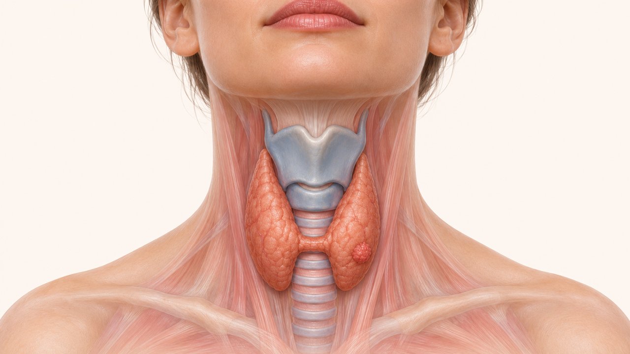
The thyroid is a butterfly-shaped endocrine gland in the midline of the neck, in front of the larynx and the windpipe. It produces thyroid hormone, which keeps organs such as the brain, heart and skeletal muscles working properly and regulates the body’s energy use; it acts as the conductor of the metabolic orchestra. When there is too little or too much thyroid hormone, many metabolic functions, growth and development can be affected, and symptoms may appear in several unrelated organs or systems. The thyroid also produces calcitonin, a hormone involved in calcium metabolism.
The thyroid and iodine
The thyroid needs iodine to work normally. Iodine is actively taken up from the bloodstream by the follicular cells that make thyroid hormone and is built into the hormone. The thyroid is the only organ in the body that stores and uses iodine.
What is a thyroid nodule?
In the broadest sense, a thyroid nodule is a growth that occupies space within the thyroid. Most are benign (not cancer), but some contain cancer. Nodules are therefore assessed with a set algorithm, so that cancer is not missed and treatment can be planned.
What are the symptoms?
A thyroid nodule may be noticed as a lump or swelling in the neck, but often it causes no symptoms and is found during an ultrasound or CT scan done for another reason. Sometimes it comes to light during tests for a high thyroid hormone level. Rarely, a nodule produces excess thyroid hormone and causes hyperthyroidism. Most nodules, including those that contain cancer, do not produce hormone. When a nodule is found, the sensible first step is therefore to measure thyroid-stimulating hormone (TSH), a pituitary hormone, to see whether the nodule is linked to excess hormone production.
When nodules are numerous and very large, they can cause neck pain, pressure on the windpipe or the oesophagus, or a feeling of something catching on swallowing. Rarely, certain fast-growing thyroid cancers or very advanced cancers present with hoarseness or with signs of skin, oesophageal or tracheal involvement.
How common are they, and what causes them?
We do not know what causes thyroid nodules, but we know they are extremely common and become more frequent with age. In Türkiye, a nodule can be detected clinically or on imaging in 50–60% of the population. Fortunately, more than 90% are benign. Nodules are more frequent in Hashimoto’s thyroiditis, the most common cause of an underactive thyroid. In some regions of Türkiye, endemic iodine deficiency leads to many nodules of varying size in the thyroid; this enlargement of the gland is known as multinodular goitre, or simply goitre.
How are thyroid nodules assessed?
When a nodule is noticed, your doctor first examines the thyroid to see whether the nodule can be felt, and to judge the number and size of nodules. Blood tests (thyroid function tests) then show whether the rest of the gland is working normally. Because examination and blood tests cannot tell whether a nodule contains cancer, a thyroid ultrasound is performed and, where needed, a fine-needle biopsy is taken.
Thyroid ultrasound
Ultrasound uses high-frequency sound waves to examine the structure and blood supply of internal organs. It involves no radiation and is easy, quick and painless, which gives it a central place in assessing thyroid nodules. With an accuracy of up to 97%, ultrasound shows whether a nodule is present, how large it is, and whether it is cystic or solid. It also guides the needle accurately into the nodule, keeping misdiagnosis and inadequate samples to a minimum.
Nodules are now grouped by risk according to how they look on ultrasound; the term “TIRADS” on your report refers to this classification. In EU-TIRADS, the European Thyroid Association’s system, category 2 means a benign appearance, 3 low risk, 4 intermediate risk and 5 high risk. The decision to biopsy weighs this risk group together with the size of the nodule: in general, biopsy is advised for nodules larger than 1 cm with a high-risk appearance, larger than 1.5 cm at intermediate risk, and larger than 2 cm at low risk. Thresholds differ somewhat between systems (for example ACR TI-RADS), and the decision is always made for your individual situation.
Fine-needle aspiration biopsy
Ultrasound-guided fine-needle biopsy is the gold standard for diagnosing thyroid nodules. The needle is very thin, so a local anaesthetic injection is usually unnecessary. Unless you take blood-thinning medication, the risk of a haematoma is generally very low, and most people return to work and normal life the same day.
The cells are examined by a cytopathologist. Results are reported in one of six categories of the Bethesda system, the common language used worldwide. The cancer risk given for each category is an average; your doctor interprets what it means for you together with the ultrasound findings.
I. Nondiagnostic
Cancer risk about 13% (5–20%)
The sample does not contain enough cells for a diagnosis; this can happen when a nodule is entirely cystic. The biopsy is usually repeated under ultrasound guidance.
II. Benign
Cancer risk about 4% (2–7%)
The most common result. If the nodule causes no cosmetic concern or pressure symptoms, surgery is not advised; it is followed with examination and ultrasound at intervals.
III. Atypia of undetermined significance (AUS)
Cancer risk about 22% (13–30%)
The cells cannot be classed as benign or malignant. Options include repeating the biopsy, molecular testing, surveillance or diagnostic surgery.
IV. Follicular neoplasm
Cancer risk about 30% (23–34%)
A needle biopsy cannot separate benign from malignant tumours in this group. Molecular testing may be used, or the thyroid lobe containing the nodule is removed to reach a diagnosis.
V. Suspicious for malignancy
Cancer risk about 74% (67–83%)
Cancer is strongly suspected, most often papillary thyroid cancer. Treatment is usually surgical.
VI. Malignant
Cancer risk about 97% (97–100%)
Cancer has been diagnosed. Treatment is usually surgical; you should see a doctor experienced in thyroid surgery.
Treatment and follow-up options
- Surveillance: Nodules shown to be benign that cause no symptoms are not operated on; they are followed with ultrasound. The biopsy may be repeated if a nodule grows or its appearance changes.
- Surgery: Used for nodules with diagnosed or suspected cancer, and for those causing pressure symptoms or cosmetic concern. Depending on the nodule, half of the thyroid (lobectomy) or the whole gland is removed.
- Non-surgical procedures: For benign nodules causing pressure symptoms or cosmetic concern, ultrasound-guided thermal ablation (radiofrequency, laser) may be an option for suitable patients who do not want surgery. A biopsy must first show that the nodule is benign. Cystic nodules can be treated with ethanol ablation.
- Active surveillance: For low-risk papillary thyroid cancers smaller than 1 cm and confined to the thyroid, close follow-up with regular ultrasound instead of immediate surgery is an accepted option in current guidelines for carefully selected patients. This decision is made together with your doctor.
What is thyroid scintigraphy, and when is it needed?
In the past, before ultrasound was used to diagnose and follow thyroid nodules, every patient had a scintigraphy scan with radioactively labelled iodine to see whether a nodule produced thyroid hormone. Nodules that do not produce hormone are called “cold”, and those that produce excess hormone “hot”. We now know that a cold nodule is not, on its own, a sign of malignancy; ultrasound and fine-needle biopsy are the gold standard for these nodules. Scintigraphy remains an important test when the thyroid is overactive. Hot nodules are unlikely to contain cancer, so a biopsy is not needed.
Sources
- Durante C, Hegedüs L, Czarniecka A, et al. 2023 European Thyroid Association Clinical Practice Guidelines for thyroid nodule management. Eur Thyroid J 2023.
- Ali SZ, Baloch ZW, Cochand-Priollet B, et al. The 2023 Bethesda System for Reporting Thyroid Cytopathology. Thyroid 2023.
- Ringel MD, Sosa JA, Baloch Z, et al. 2025 American Thyroid Association Management Guidelines for Adult Patients with Differentiated Thyroid Cancer. Thyroid 2025.
- Papini E, Monpeyssen H, Frasoldati A, Hegedüs L. 2020 European Thyroid Association Clinical Practice Guideline for the Use of Image-Guided Ablation in Benign Thyroid Nodules. Eur Thyroid J 2020.