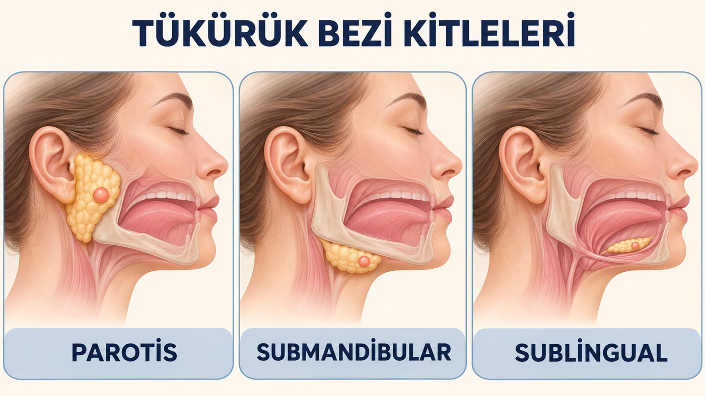
In brief
- Salivary gland cancers are rare diseases that arise from malignant cells in the tissues of the salivary glands.
- Exposure to radiation increases the risk of salivary gland cancer.
- The main symptoms are a swelling in the neck and difficulty swallowing.
- Examination of the head, neck and mouth is the basis of diagnosis.
- Certain factors affect the chance of recovery (prognosis) and the treatment options.
The salivary glands
The salivary glands make saliva and release it into the mouth. Saliva contains enzymes that help digest food and antibodies that protect against infections in the mouth. There are three pairs of large (major) salivary glands:
- Parotid glands: The largest salivary glands, lying just in front of and below each ear. Most salivary gland tumours arise here.
- Sublingual glands: In the floor of the mouth, under the tongue.
- Submandibular glands: Just below the jawbone.
There are also hundreds of small (minor) salivary glands in the mouth, nose and larynx that can be seen only under a microscope. Most minor salivary gland tumours start in the palate.
More than half of salivary gland tumours are benign and do not spread to other tissues. Salivary gland cancer is a type of head and neck cancer.
Risk factors
Anything that increases the chance of getting a disease is called a risk factor. Having a risk factor does not mean you will get cancer, and cancer can develop without one. Talk to your doctor if you think you may be at risk. The causes of most salivary gland cancers are unknown, but older age, previous radiotherapy to the head and neck, and exposure to certain substances at work are known risk factors.
Symptoms
Salivary gland cancer may cause no symptoms; a tumour may be found during a routine dental check, a health check or a physical examination. See your doctor if you have any of the following:
- A lump, usually painless, in the ear, cheek, jaw, lip or inside the mouth
- Fluid draining from the ear
- Difficulty swallowing or opening the mouth wide
- Numbness or weakness in the face
- Pain in the face that does not go away
These signs and symptoms may be due to salivary gland cancer or to other conditions.
Methods of diagnosis
One or more of the following may be used:
- History and physical examination: The body, and especially the head and neck, is examined for lumps and anything unusual. The patient’s general health, habits, past illnesses and treatments are reviewed. The parotid and submandibular glands are examined by hand, and the salivary duct by a method called bimanual examination.
- Magnetic resonance imaging (MRI): Detailed images of the area are obtained using a magnet, radio waves and a computer.
- Computed tomography (CT): Detailed cross-sectional images are created using X-rays. Contrast material given into a vein or swallowed makes blood vessels and tumours clearer and easier to tell apart from surrounding tissue.
- PET scan (positron emission tomography): Finds cancer cells in the body. A small amount of glucose (sugar) carrying a radioactive label is given into a vein and the scanner shows where glucose is being used. Cancer cells use more glucose and so appear brighter.
- Endoscopy: The mouth, throat and larynx are examined through the mouth with a thin, tube-like instrument that has a light and a lens. A special method called sialendoscopy is used to diagnose and treat problems in the salivary duct such as stones, strictures and plugs.
- Biopsy: Cells or tissue are removed and examined under the microscope by a pathologist.
- Fine-needle aspiration biopsy (FNA): A sample of tissue or fluid is taken with a very thin needle and examined. It is the most commonly used type of biopsy for salivary gland cancer.
I can feel a lump in my cheek; could it be cancer?
When a lump is noticed in the cheek, the first step is to ask how it appeared. In someone past middle age who has long used alcohol or tobacco, a lump accompanied by pain, numbness of the facial muscles, limited jaw movement and an enlarged lymph node in the neck is more likely to be cancer.
A swelling that comes on suddenly, appears with cramp-like pain during meals and may come and go is, on the other hand, more likely to be caused by a stone or inflammation in the salivary glands.
Salivary gland cancer
How common is it?
Salivary gland tumours are rare, with about 2.5–3 cases per 100,000 people each year. They make up about 3–5% of head and neck cancers. Their microscopic appearance and clinical behaviour vary so widely that diagnosis and treatment can be very challenging. Most patients with malignant salivary gland tumours are in their fifties and sixties.
Risk factors
Exposure to ionising radiation is the factor most often implicated in salivary gland cancer. The risk is also raised by exposure in rubber industry workers, miners exposed to asbestos, plumbers and people who work with timber.
Tumour types
Under the microscope, salivary gland tumours are the most varied of all tumour groups in the body. The World Health Organization classification, updated in 2022, defines a large number of benign and malignant tumour types, and the number grows with each edition; some of these types are very rare. Correctly identifying the type and grade of the tumour directly affects the choice of treatment. The most common benign tumour is pleomorphic adenoma, which makes up 50% of all salivary gland tumours and 65% of all parotid tumours. The most common malignant tumour is mucoepidermoid carcinoma, which makes up 10% of all salivary gland tumours and 35% of malignant ones. It too arises most often in the parotid.
Clinical features
Most patients with a benign tumour present with a slowly growing, painless swelling in a salivary gland. In malignant tumours, a rapidly growing mass may be accompanied by numbness or weakness from involvement of the facial nerve, pain, a lump in the neck, and redness and hardening of the skin over the mass.
Factors affecting outcome and the choice of treatment
- The stage of the cancer (especially the size of the tumour)
- Which salivary gland the cancer is in
- How different the cancer cells look from normal tissue under the microscope
- The patient’s age and general condition
Treatment
Treatment of salivary gland tumours is planned according to the type, grade, site and stage of the tumour.
Benign tumours
Benign tumours are treated with surgery. The tumour is removed together with a margin of healthy gland tissue. Pleomorphic adenoma, the most common benign tumour, is generally removed because it can grow over time and, rarely, turn malignant.
Malignant tumours
- Surgery: Surgery is the mainstay of treatment for salivary gland cancer. For tumours of the parotid gland, part or all of the gland is removed (parotidectomy). The facial nerve, which moves the muscles of the face, runs through the parotid gland, so finding and preserving it is essential; nerve monitoring is used during the operation for this purpose. For tumours of the submandibular gland, the whole gland is removed; minor salivary gland tumours are removed with a margin of healthy tissue.
- Neck dissection: If the cancer has spread to the neck lymph nodes, or the tumour is high grade or advanced, the lymph nodes in the neck are also cleared.
- Radiotherapy: Added after surgery for high-grade or advanced tumours, when the tumour could not be removed completely, or when nerves or lymph nodes are found to be involved.
- Drug treatments: For disease that cannot be removed surgically or has spread to distant organs, chemotherapy and targeted drugs chosen according to the molecular features of the tumour may be used.
Swellings caused by stones and inflammation
In suitable patients, stones and strictures in the salivary duct can be treated from inside the duct by sialendoscopy, without removing the gland.
Sources
- Skálová A, Hyrcza MD, Leivo I. Update from the 5th Edition of the World Health Organization Classification of Head and Neck Tumors: Salivary Glands. Head and Neck Pathology 2022.
- Geiger JL, Ismaila N, Beadle B, et al. Management of Salivary Gland Malignancy: ASCO Guideline. Journal of Clinical Oncology 2021.
- Canadian Cancer Society. Surgery for salivary gland cancer.